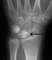
The cause of Kienböck’s disease unknown. Many patients with Kienböck’s disease think they have a sprained wrist and in many cases there is a prolonged period of time until they are correctly diagnosed. In Kienböck’s disease, the blood supply to the lunate, one of the small bones of the hand near the wrist is interrupted and the bone undergoes changes and eventually collapses.
Patients often complain of a painful and sometimes swollen wrist. there may be a limited range of motion in the affected wrist (stiffness) and decreased grip strength in the hand. Often there can be tenderness directly over top of the wrist and pain or difficulty in turning the hand upward.
Kienböck’s disease progresses through four stages. In its early stages, Kienböck’s disease may be difficult to diagnose because the symptoms are so similar to those of a sprained wrist. Even X-rays of the wrist may appear normal.
Stage 1: X-rays may be normal or suggest a possible fracture. Magnetic resonance imaging (MRI) may also be helpful in making the diagnosis in this early stage.

Stage 2: The lunate bone begins to harden. Brighter or whiter areas on X-rays indicate that the bone is dying. MRI or computed tomography (CT) may be used to assess the bone. Wrist pain, swelling, and tenderness are common.

Stage 3: The dead bone begins to collapse and break into pieces. As the bone begins to break apart, the surrounding bones may begin to shift position. Increasing pain, weakness in gripping, and limited motion may be experienced.

Advanced Grade 3B Kienbock’s disease with lunate collapse
Stage 4: The surfaces of adjoining bones are affected. One result may be generalised arthritis of the wrist.
Although there is no absolute cure, there are several nonsurgical and surgical options for treating this disease. The treatment depends on at which stage the lunate has reached. The goals of treatment are to relieve the pressure on the lunate and to try to restore blood flow within the bone.
Early on the wrist may be splinted or casted for two to three weeks. Anti-inflammatory medications, such as aspirin or ibuprofen, will help relieve any pain and reduce swelling.
There are several surgical options for treating the more-advanced stages of Kienböck’s disease. The choice of procedure will depend on several factors, including disease progression, activity level, personal goals, and the surgeon’s experience with the procedures.
In some cases, it may be possible to return the blood supply to the bone (revascularization). This procedure takes portion of bone (graft) from the inner bone of the lower arm.
If the bones of the lower arm are uneven in length, a joint leveling procedure may be recommended. The radius bone may be shortened by removing a section of the bone. This leveling procedure reduces the forces that bear down on (compress) the lunate and seems to halt progression of the disease.
If the lunate is severely collapsed or fragmented into pieces, it can be removed with the two bones on either side of the lunate. This procedure, called a proximal row carpectomy (PRC), will relieve pain while maintaining partial wrist motion.
Another procedure that eases pressure on the bone is fusion. In this procedure, several of the small bones of the hand are fused together. If the disease has progressed to severe arthritis of the wrist, fusing the bones will reduce pain and help maintain function. The range of wrist motion, however, will be limited.
There is also the option in certain cases of undertaking a lunate replacement.
Mr Sorene has an interest in Kienbock’s disease and undertakes all of the most modern treatments for Kienbock’s disease including arthroscopic treatment, wrist denervation, limited wrist fusions, proximal row carpectomy and lunate replacement surgery.
Wrist arthroscopy – ‘Keyhole surgery’

Proximal Row Carpectomy (PRC)

Lunate Replacement
